
| Origin | Mouse | |
| Antibody | ||
| Nomenclature | ||
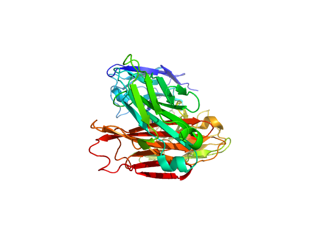
| PDB Code | 2R2E | |
| Resolution (Å) | 2.4 | |
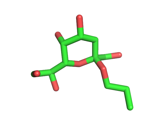
| Reference | Brooks, C.L., Muller-Loennies, S., Brade, L., Kosma, P., Hirama, T., MacKenzie, C.R., Brade, H., Evans, S.V. Exploration of specificity in germline monoclonal antibody recognition of a range of natural and synthetic epitopes. Primary Citation Exploration of specificity in germline monoclonal antibody recognition of a range of natural and synthetic epitopes. Brooks, C.L., Muller-Loennies, S., Brade, L., Kosma, P., Hirama, T., MacKenzie, C.R., Brade, H., Evans, S.V. Journal: (2008) J.Mol.Biol. 377: 450-468 PubMed: 18272175 DOI: 10.1016/j.jmb.2008.01.018 Search Related Articles in PubMed PubMed Abstract: To explore the molecular basis of antigen recognition by germline antibodies, we have determined to high resolution the structures of the near-germline monoclonal antibody S25-2 in complex with seven distinct carbohydrate antigens based on the bacterial sugar 3-deoxy-alpha-D-manno-oct-2-ulosonic acid (Kdo). In contrast to previous findings, the inherited germline Kdo monosaccharide binding site is not restricted to this bacterial sugar but is able to accommodate an array of substitutions and chemical modifications of Kdo, including naturally occurring antigens containing the related monosaccharide d-glycero-alpha-d-talo-oct-2-ulosonic acid as well as nonterminal Kdo residues. However, we show by surface plasmon resonance and ELISA how antibody S25-2 specificity is so dependent on the context in which the antigen is presented that a free disaccharide displays strong binding while the same lipid-A-bound disaccharide does not bind. These structures provide insight into how inherited germline genes code for immunoglobulins of limited flexibility that are capable of binding a range of epitopes from which affinity-matured antibodies are generated. Keywords: Animals, Antibodies, Monoclonal, Antibody Specificity, Antigens, Biological Products, Chlamydophila psittaci, Enzyme-Linked Immunosorbent Assay, Epitopes, Lipopolysaccharides, Mice, Models, Molecular, Molecular Structure, Protein Binding, Protein Structure, Tertiary, Sugar Acids, Surface Plasmon Resonance Related Structures: Primary Citation of: 2R1W 2R1X 2R1Y 2R23 2R2B 2R2E 2R2H 3BPC Organizational Affiliation: Department of Biochemistry and Microbiology, University of Victoria, PO Box 3055 STN CSC, Victoria, BC, Canada. Click on abstract words and keywords to add them to the search box. [Hide Abstract] Molecular Description Hide Classification: Immune System Structure Weight: 48632.46 Molecule: Fab, antibody fragment (IgG1k), light chain Polymer: 1 Type: protein Length: 219 Chains: A Organism: Mus musculus Molecule: Fab, antibody fragment (IgG1k), heavy chain Polymer: 2 Type: protein Length: 222 Chains: B Organism: Mus musculus Structure Validation Hide View the full validation report MolProbity Ramachandran Plot Download Ramachandran Plot PDF (from MolProbity ) Source Hide Polymer: 1 Scientific Name: Mus musculus Taxonomy Common Name: Mouse Polymer: 2 Scientific Name: Mus musculus Taxonomy Common Name: Mouse Related PDB Entries Hide Identifier Details 1Q9Q The same protein with a different ligand 2R1W 2R1X 2R1Y 2R23 2R2B 2R2H Ligand Chemical Component Hide Identifier Formula Name View Interactions KDE Search Download KDE C11 H18 O8 prop-2-en-1-yl 3-deoxy-beta-L-gulo-oct-2- ulopyranosidonic acid KDE:2R2E Ligand Explorer Jmol External Domain Annotations Hide CATH Classification v4.0.0: 4 Domains – data from CATH Pfam Classification: 6 Domains – data from Pfam Data in orange boxes are gathered from external resources (when available). Reset Layout, (2008), 10.2210/pdb2r2e/pdb | |
💡 Mouse: rotation | Scroll wheel: zoom
Styles: